Gallery
BOMP image competition 2022
Winner of 'Top 3 images'
-
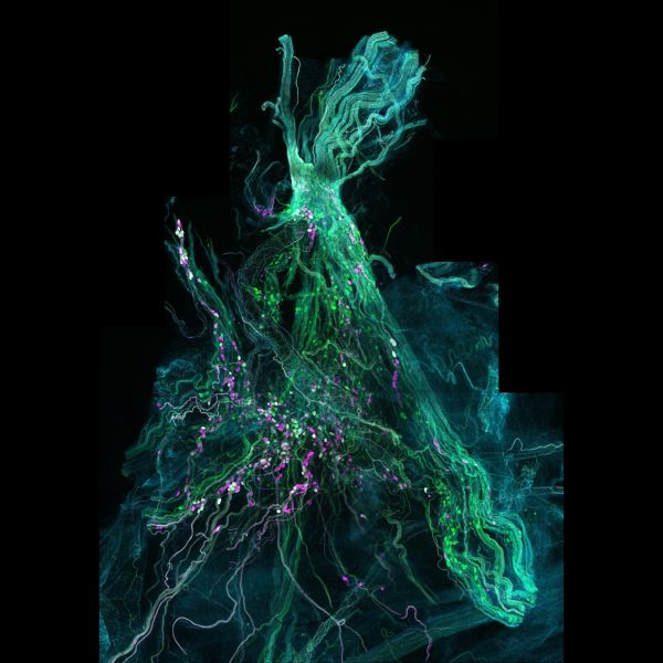
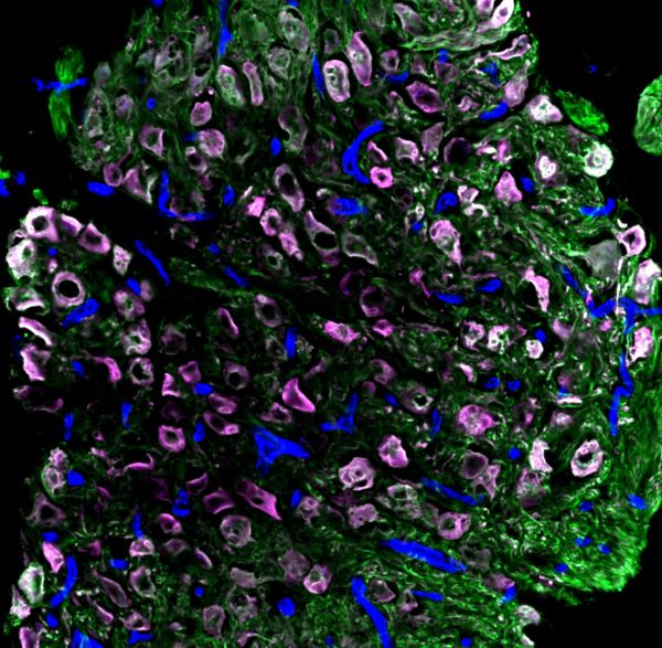
Mighty pelvic ganglion by John-Paul Fuller-Jackson, LSM900
Title: Mighty pelvic ganglion by John-Paul Fuller-Jackson Description: The major pelvic ganglion of the rat showing the various populations of neurons that reside within it. The nerves coming out of this ganglion are critical for pelvic organ function. Instrument:LSM900 -
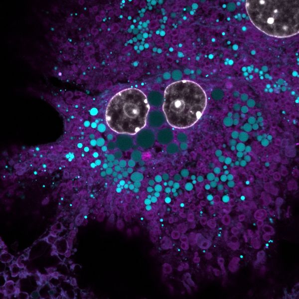
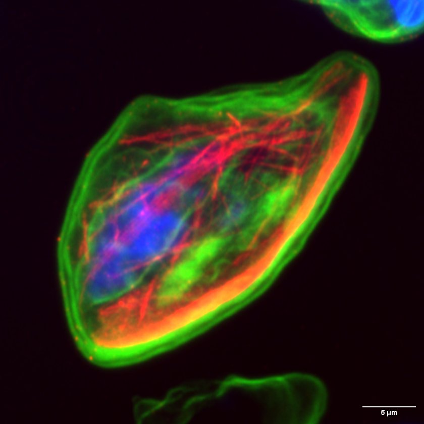
Smilin' cell-fie by Stacey Keenan, LSM900
Title: Smilin' cell-fie by Stacey Keenan, Description: Primary mouse hepatocyte stained with DAPI, BODIPY and MitoTracker to visualise nucleus, lipid droplets and mitochondria Instrument: LSM900 -
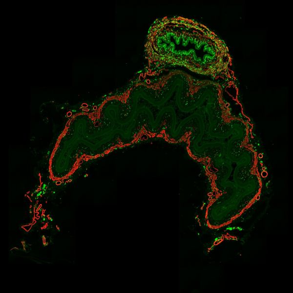
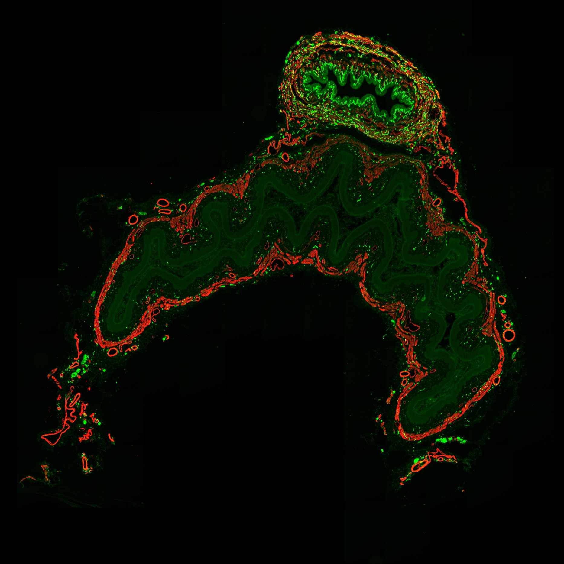
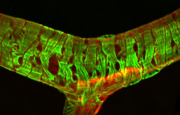
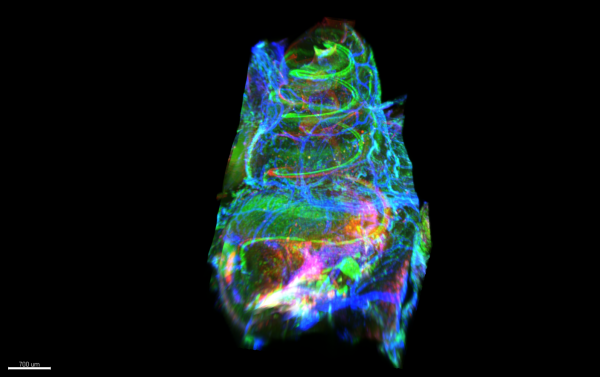
Unconventional Christmas Tree by Jonathan Chan, Axioscan7
Title: Unconventional Christmas Tree by Jonathan Chan Description: Christmas tree with red and green lights, plus a round shining star at the top. Transverse section of rat lower abdomen stained for CGRP axons (green), smooth muscle (red). Instrument: Axioscan 7


Note: Image size was reduced for some images.
Submitted entries
-
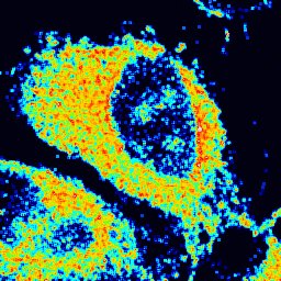
FLIM HeLa cell by Soheila Sabouri, FV3000
Title: FLIM HeLa cell by Soheila Sabouri Description: It is a FLIM result of HeLa cells stained by new generation of fluorescent probes (AIE: Aggregation-Induced Emission). Lifetme of the dye in every part of cell is different which shows the combination of this AIE dye and the imaging technique is successful to map and measure intracellular microenvironment accurately. Instrument:FV3000 -
Anisotropy FAIM by Soheila Sabouri, FV3000
Title: Anisotropy FAIM by Soheila Sabouri Description: Fluorescence Anisotropy Imaging microscopy of HeLa cells stained by new generation of fluorophores (AIE: aggregation-induced emission). Instrument:FV3000 -

Neuromuscular Junction by Chris Karagiannis, LSM900 Airyscan
Title: Neuromuscular Junction by Chris Karagiannis Description: This is a composite image of a neuromuscular junction. Every muscle fiber is innervated by a single neuron and at the intersection of this innervation forms the displayed junction. Red: microtubules following the axonal body. Blue: synaptic vesicles localized to the nerve terminal. Green: acetylcholine receptors at the muscle endplate. Instrument:LSM900 Airyscan -
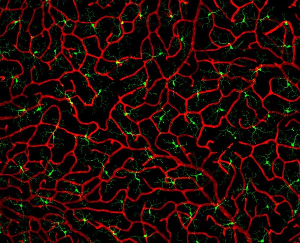
Bandaged vessel by Pialuisa Quiriconi, LSM800
Title: Bandaged vessel by Pialuisa Quiriconi, LSM800 Description: The retina is the light-sensitive tissue sitting at the back of the eye and is responsible for vision. This image depicts a large blood vessel (red) in the retina and the contractile cells (green) wrapping around it like bandage, that control vessel tone to regulate blood flow. -
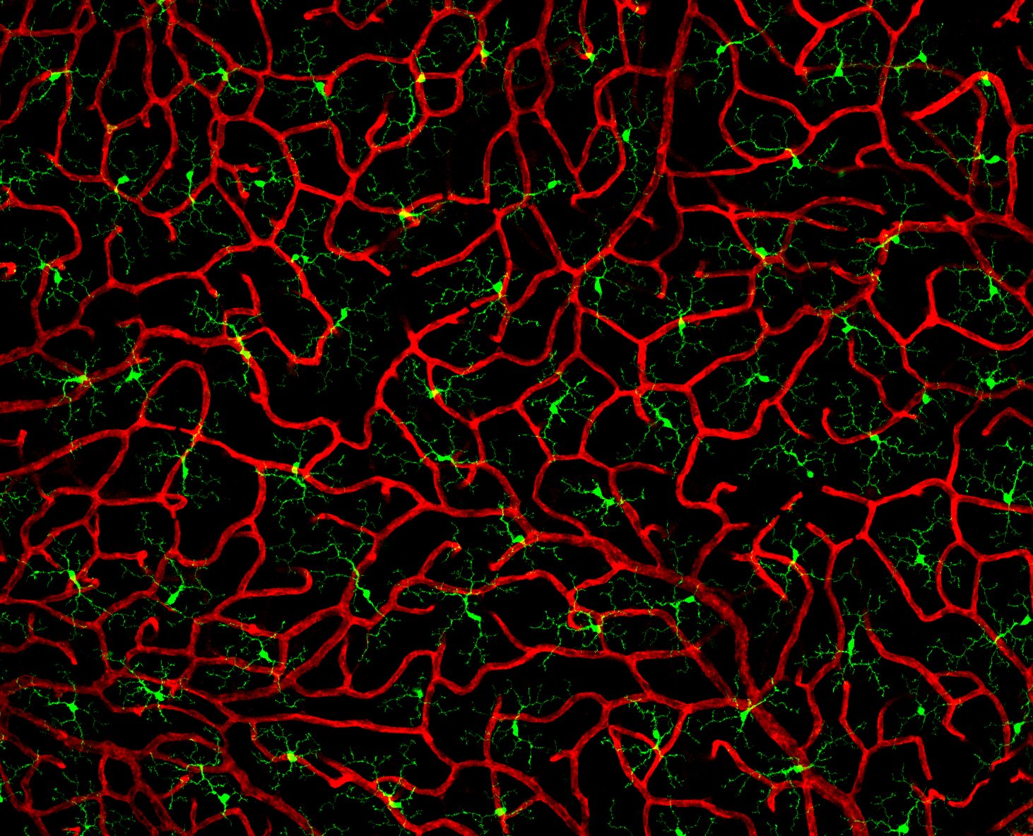
Coral in the eye by Pialuisa Quiriconi, LSM800
Title: Coral in the eye by Pialuisa Quiriconi, LSM800 Description: The retina is the light-sensitive tissue sitting at the back of the eye and is responsible for vision. This image depicts the finest blood vessels (red) in the retina called capillaries. Many cells reside amongst this dense network of vessels that resembles coral, including immune cells called microglia (green). -
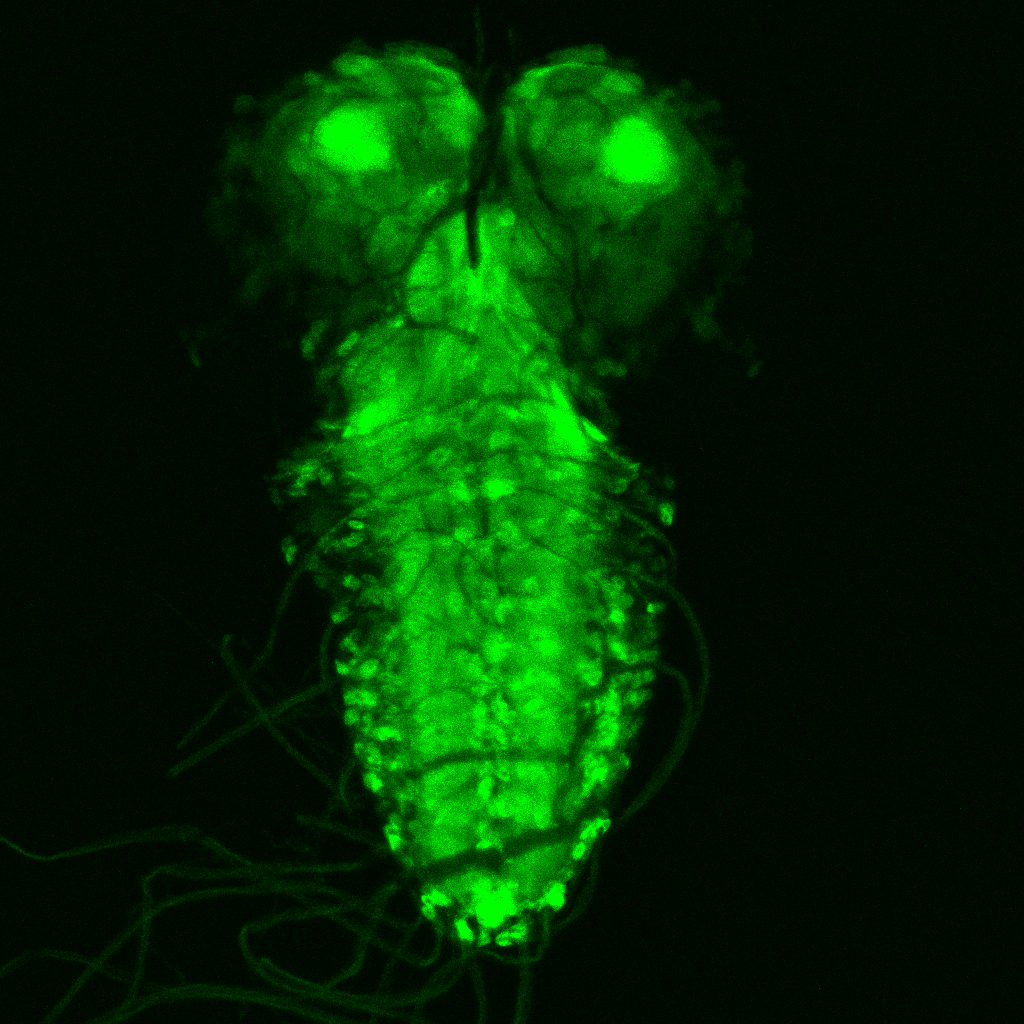
Gaze of the abyss by Wei Chen, Leica SP8
Title: Gaze of the abyss by Wei Chen, Leica SP8 Description: The image shows the expression of an important neuronal receptor from Drosophila larval brain, that is targeted by the most widely used insecticide imidalorpid, which provide better understanding towards the insecticide's mode of action and help improve the design of insecticides of the future. -
Neuromuscular Junction by Chris Karagiannis, LMS900
Title: Neuromuscular Junction by Chris Karagiannis, LMS900 Description: The intersection between a neuron and the muscle fiber being innervated forms the imaged neuromuscular junction. -
The Ghost by Chau Le Bao Tran, LSM900
Title: The Ghost by Chau Le Bao Tran, LSM900 Description: Transplanted human cells (cell body in white and human nucleus in red) in mouse brain (DAPI in blue). Important for drug screening on human tissue in living host -
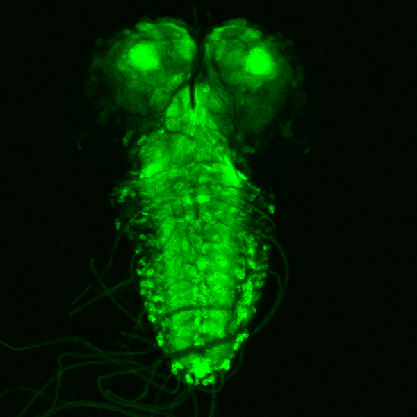
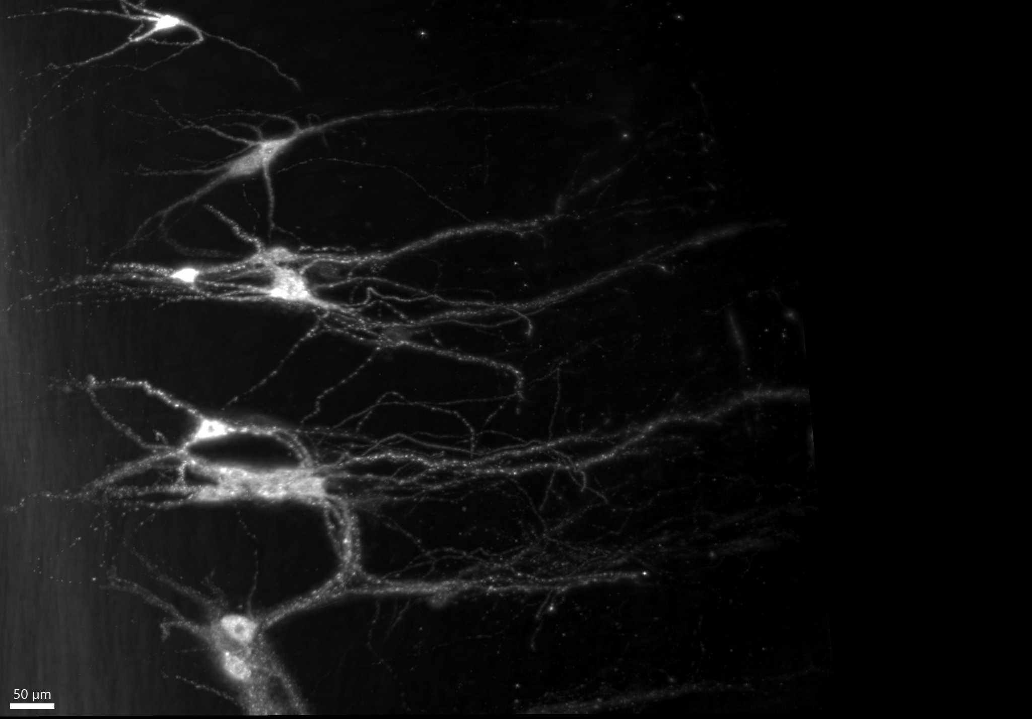
A few motoneurons by John-Paul Fuller-Jackson, Ultramicroscope II
Title: A few motoneurons by John-Paul Fuller-Jackson, Ultramicroscope II Description: Skeletal muscle is controlled by motoneurons in the spinal cord, which integrate signals from the environment and the brain via the spread of fibers seen in the image known as dendrites. -

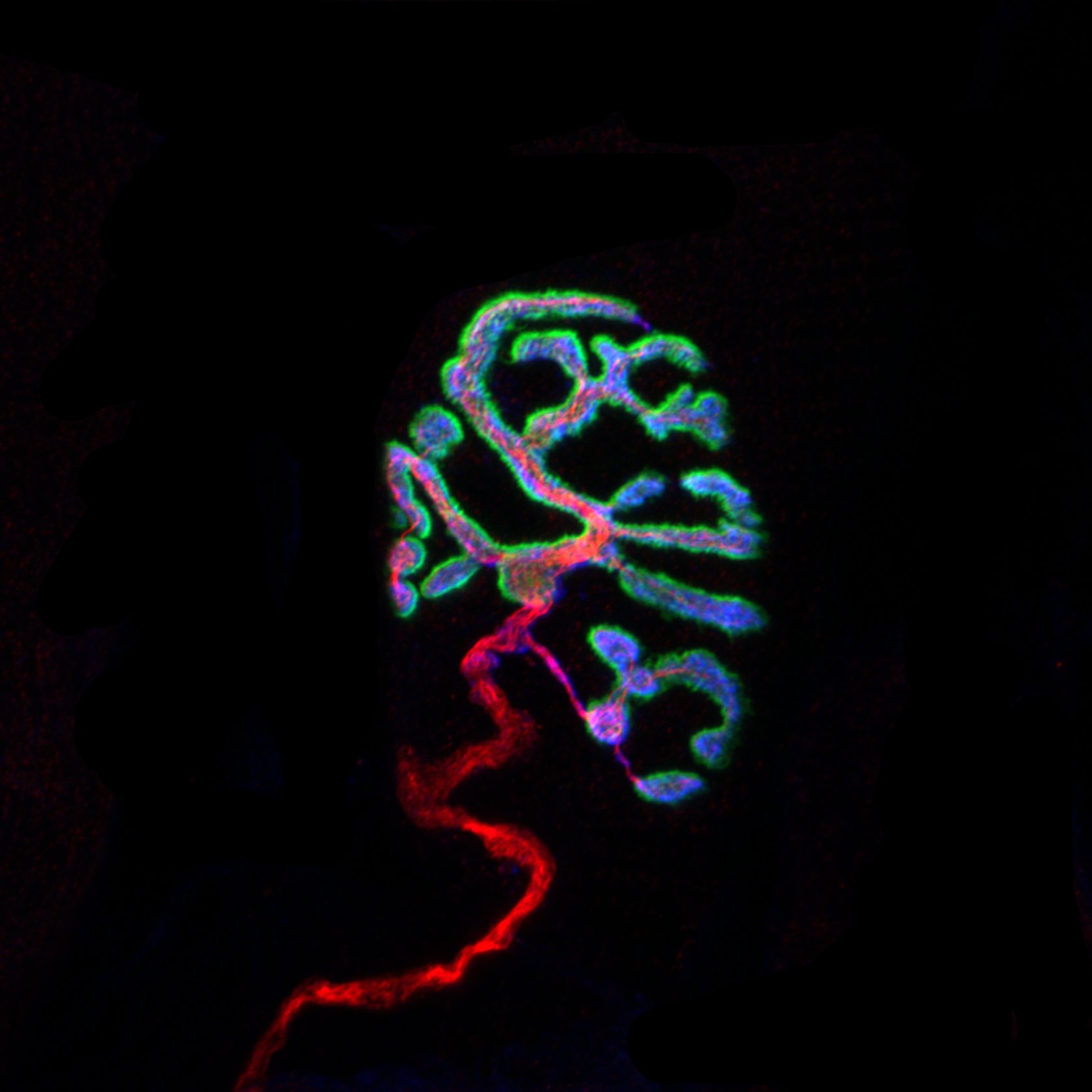
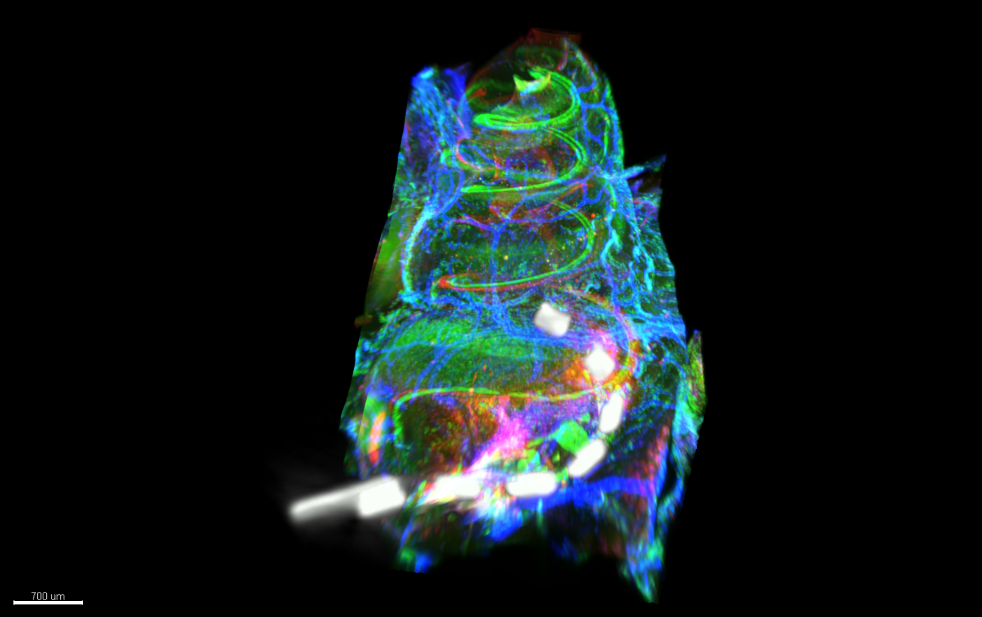
Cochlear Implant in situ by Kate Brody, Ultramicroscope II
Title: Cochlear Implant in situ by Kate Brody, Ultramicroscope II Description: Light sheet microscopy image showing a cochlea which is implanted with a cochlear implant. Hair cells and macrophages are labelled in the green, neurons and extracellular matrix in the red and arterioles and myo-fibroblasts in the blue. Co-registered with the image is a micro-CT of the cochlear implant in situ -
Cochlea in colour by Kate Brody, Ultramicroscope II
Title: Cochlea in colour by Kate Brody, Ultramicroscope II Description: Light sheet microscopy image showing a cochlea which is implanted with a cochlear implant. Hair cells and macrophages are labelled in the green, neurons and extracellular matrix in the red and arterioles and myo-fibroblasts in the blue. -
Plasmodium falciparum gametocyte microtubules by Jiahong Li, Elyra/LSM880
Title: Plasmodium falciparum gametocyte microtubules by Jiahong Li, Elyra/LSM880 Description: The unusual nuclear microtubules appear in the non-mitotic stage Plasmodium falciparum gametocyte. Understand the structure and function of nuclear microtubules can help us to block gametocyte development and malaria diseases transmission. The microtubules were labeled with anti-beta tubulin (red), the chromatins were labeled with DAPI, and the general proteins were stained with NHS-ester 488 dye. -
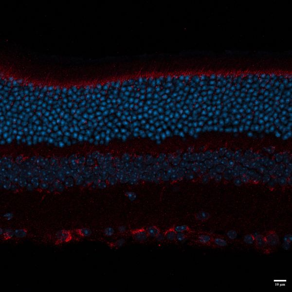
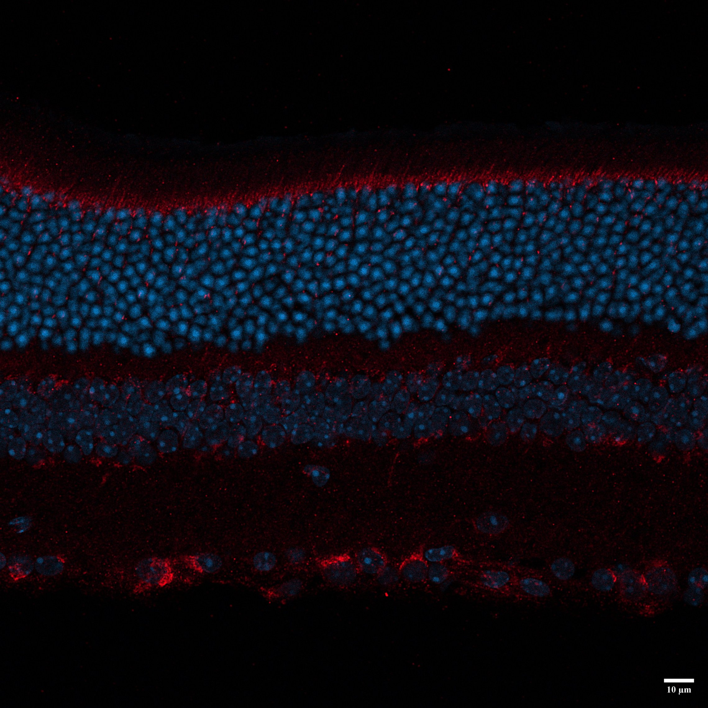
BLACK MOUSE P35 by Nadia Hosseini Naveh, LSM800
Title: BLACK MOUSE P35 by Nadia Hosseini Naveh, LSM800 Description: this image shows different layers of retina , I used LAMP1 for primary antibody , and 647 and DAPI for secondary . -
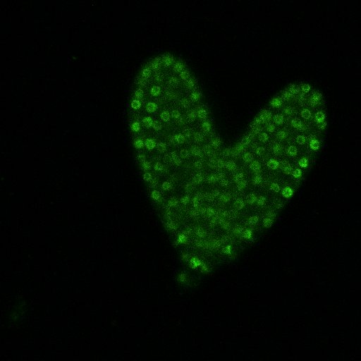
I 'Heart' Microscopy by Nicole Thomson, Nikon C2
Title: I 'Heart' Microscopy by Nicole Thomson, Nikon C2 Description: Arabidopsis thaliala heart stage embryo with gLUH nuclei expression -
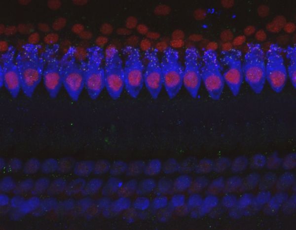
Synaptic puncta on Hair Cells in the cochlea by Alexandra Hill, Nikon A1R
Title: Synaptic puncta on Hair Cells in the cochlea by Alexandra Hill, Nikon A1R Description: Here is a 60x coloc image of the IHC of a Cat cochlea, this was taken from a frequency point of 12kHz on a normal hearing cochlea. you can see the inner Hair cells i quiet vibrant and show Lots of synaptic Puncta on each cell. -
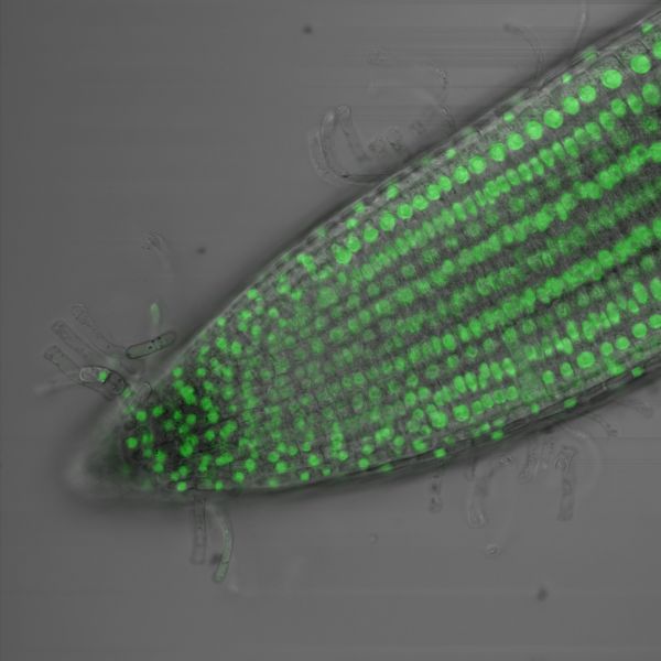
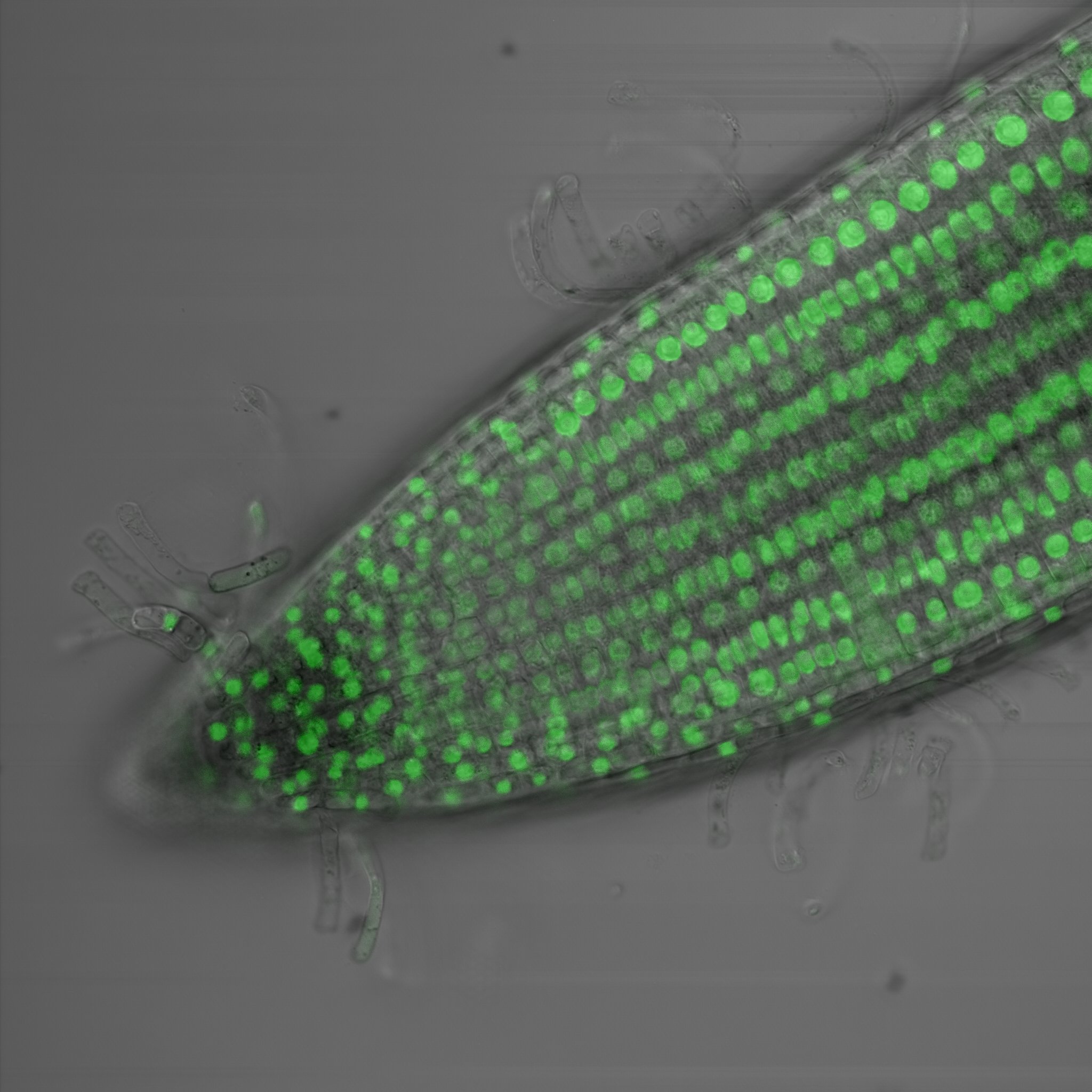
Green fluorescence in the root tip of Arabidopsis thaliana by Amelia Keynton, Nikon C2
Title: Green fluorescence in the root tip of Arabidopsis thaliana by Amelia Keynton, Nikon C2 Description: The green dots which represent the nucleus of the cell tell us that our fluorescent protein is being expressed in every cell in the root tip of Arabidopsis thaliana. We use this to determine where specifically in the root different genetic elements called promoters can drive protein expression. -
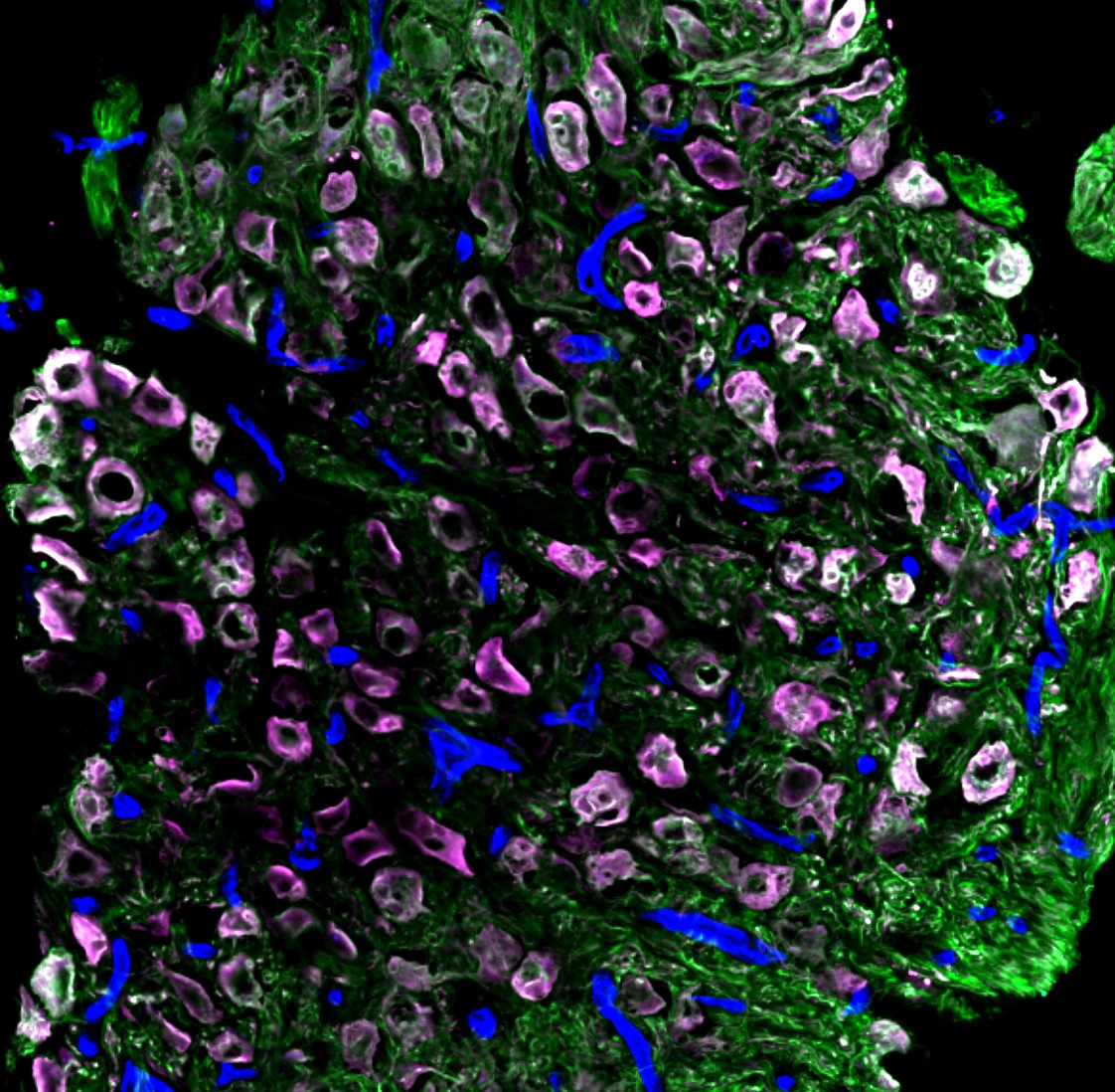
Peacock ganglia by Sapna Devi, Zeiss LSM780
Title: Peacock ganglia by Sapna Devi, Zeiss LSM780 Description: A collection of neurons and blood vessels key to functioning of the nervous system -
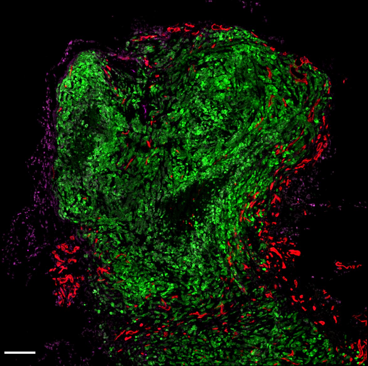
Green Alien by Sapna Devi, Zeiss LSM780
Title: Green Alien by Sapna Devi, Zeiss LSM780 Description: Tumour cells in green invading breast tissues surrounded by formation of new blood cells in red. Image shows how tumours expand and take over a normal tissue quickly. -
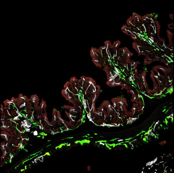
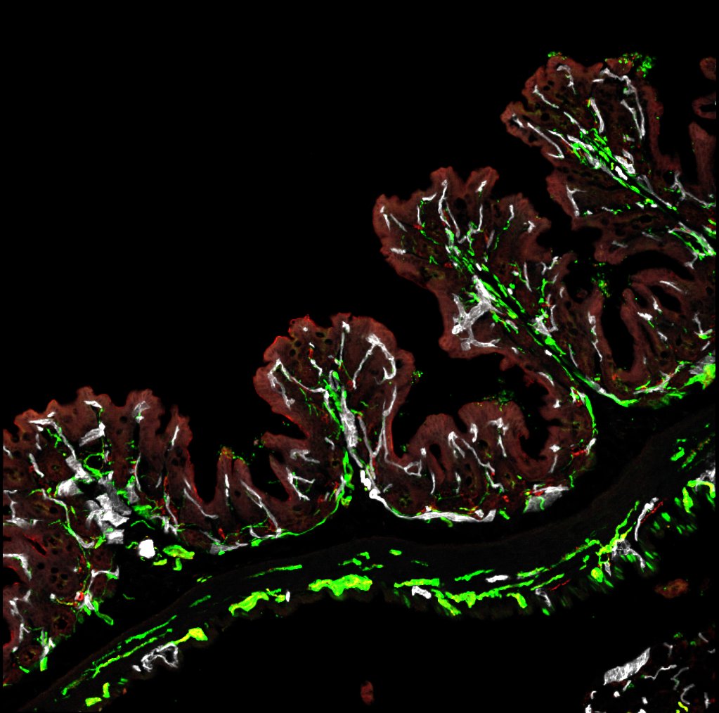
Ferny gut by Sapna Devi , Zeiss LSM780
Title: Ferny gut by Sapna Devi , Zeiss LSM780 Description: A typical structure found in the colon with lots of nerve innervation in green colocalizing with blood vessels in white. Image demonstrates how the body "talks" to the gut. -
Traveling thru space by Sapna Devi, Zeiss LSM780
Title: Traveling thru space by Sapna Devi, Zeiss LSM780 Description: Cells known as fibroblastic reticular cells key in tissue architecture. -
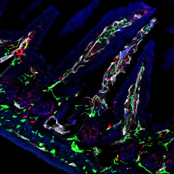
Gut feelings by Sapna Devi, Zeiss LSM780
Title: Gut feelings by Sapna Devi, Zeiss LSM780 Description: Nerves associated with blood vessels seen in the villus of the small intestine crucial for controlling gut functions. -
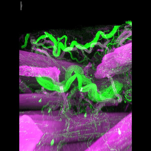
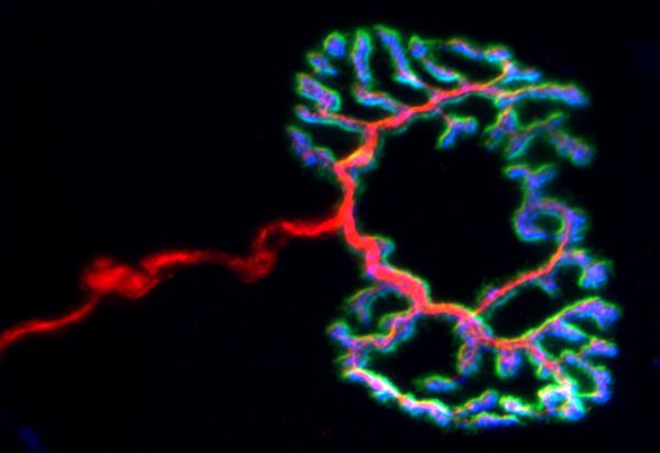
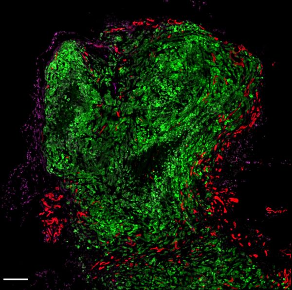
Nervous Break-in by Jonathan Chan, UMII
Title: Nervous Break-in by Jonathan Chan, UMII Description: A big axon (green) weaves in between a break in the smooth muscle layer (purple) in bladder serosa. A gentle reminder to take a well-deserved break when even when you have a wall of activities. -
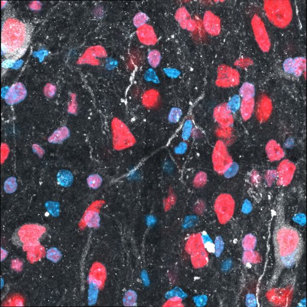
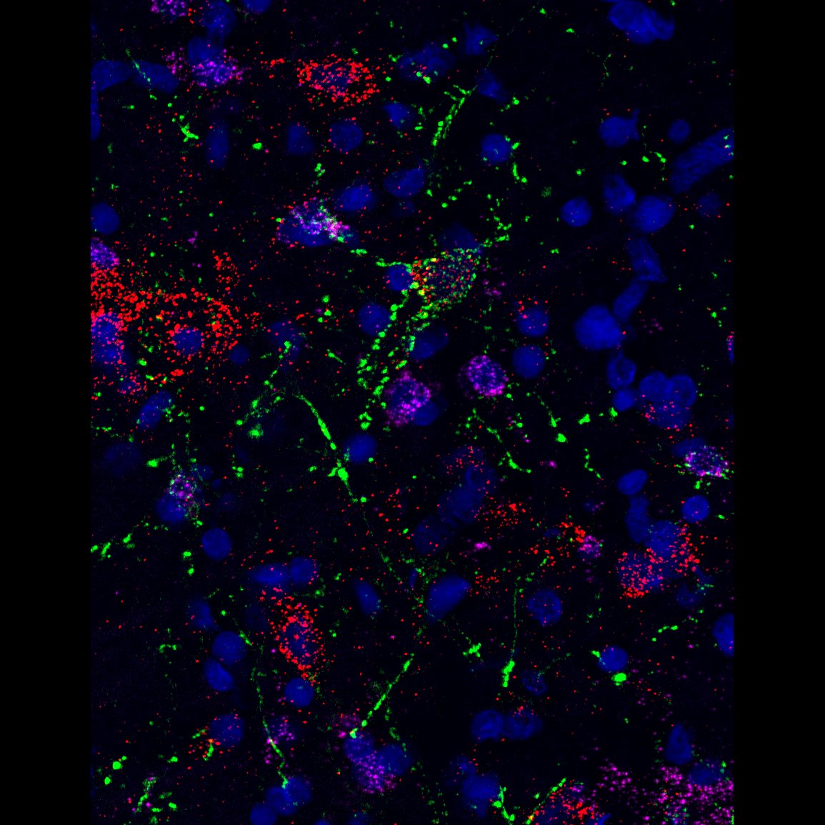
dot-to-dot by Angela Connelly, LSM880
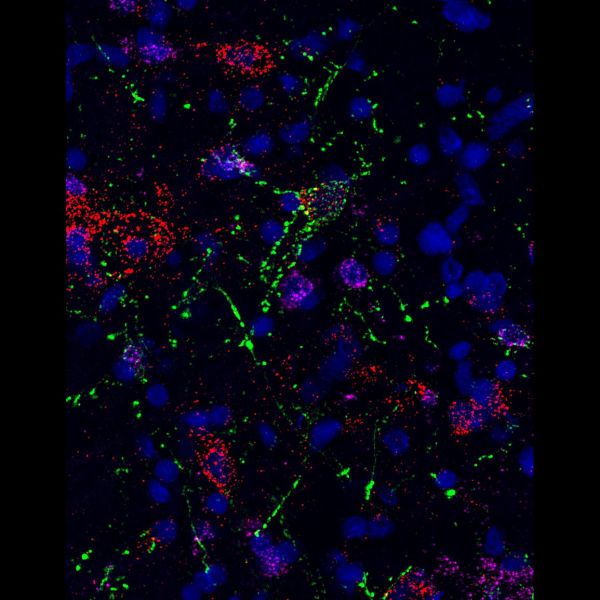
Title: dot-to-dot by Angela Connelly, LSM880 Description: RNAscope image of the preBotzinger complex in the rat brainstem, with VGlut (Cy5), VGAT (Cy3) and GtACR2 opsin photoinhibited neurons in green. The neurochemistry of preBotzinger neurons which enable and coordinate breathing; which is vital for life, and have been inhibited via optogenetics, is great to understand. -
Well, well, well what do we have here? by Teagan Wagner, Olympus FVMPERS 2-Photon
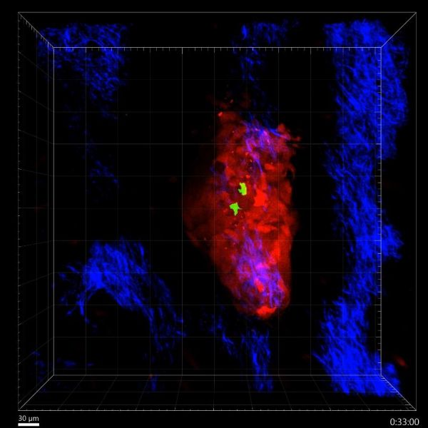
Title: Well, well, well what do we have here?' T cells survey melanoma in the skin by Teagan Wagner, Olympus FVMPERS 2-Photon Microscope Description: Specialist immune cells, known as T cells, are important for recognising and fighting cancer. This video shows T cells (green) actively recognising and surveying a melanoma tumour (red) in the skin (blue) in 3-D in real-time for more than 2 hours. Video: https://youtu.be/zXcrkh908oI


Note: Image size was reduced for some images.